

Imaging & Radiology Services
Schedule Imaging & Radiology Services
Explore available imaging services, find a location, and schedule your appointment online.
Central Valley Doctors Health System uses radiology imaging technology and offers all modalities of radiological services to help you and your doctor pinpoint the cause of your symptoms. Our team of radiologists is committed to your satisfaction throughout our multiple Modesto & Turlock locations.

Our Imaging Services
- Computed Tomography (CT) scans
- Magnetic Resonance Imaging (MRI)
- High-definition breast MRI
- Ultrasound imaging
- X-ray services
- Bone densitometry (DXA) exams
- Nuclear medicine tests
- And much more
Find an Imaging Center in Modesto & Turlock

Doctors Hospital of Manteca

Doctors Medical Center Modesto
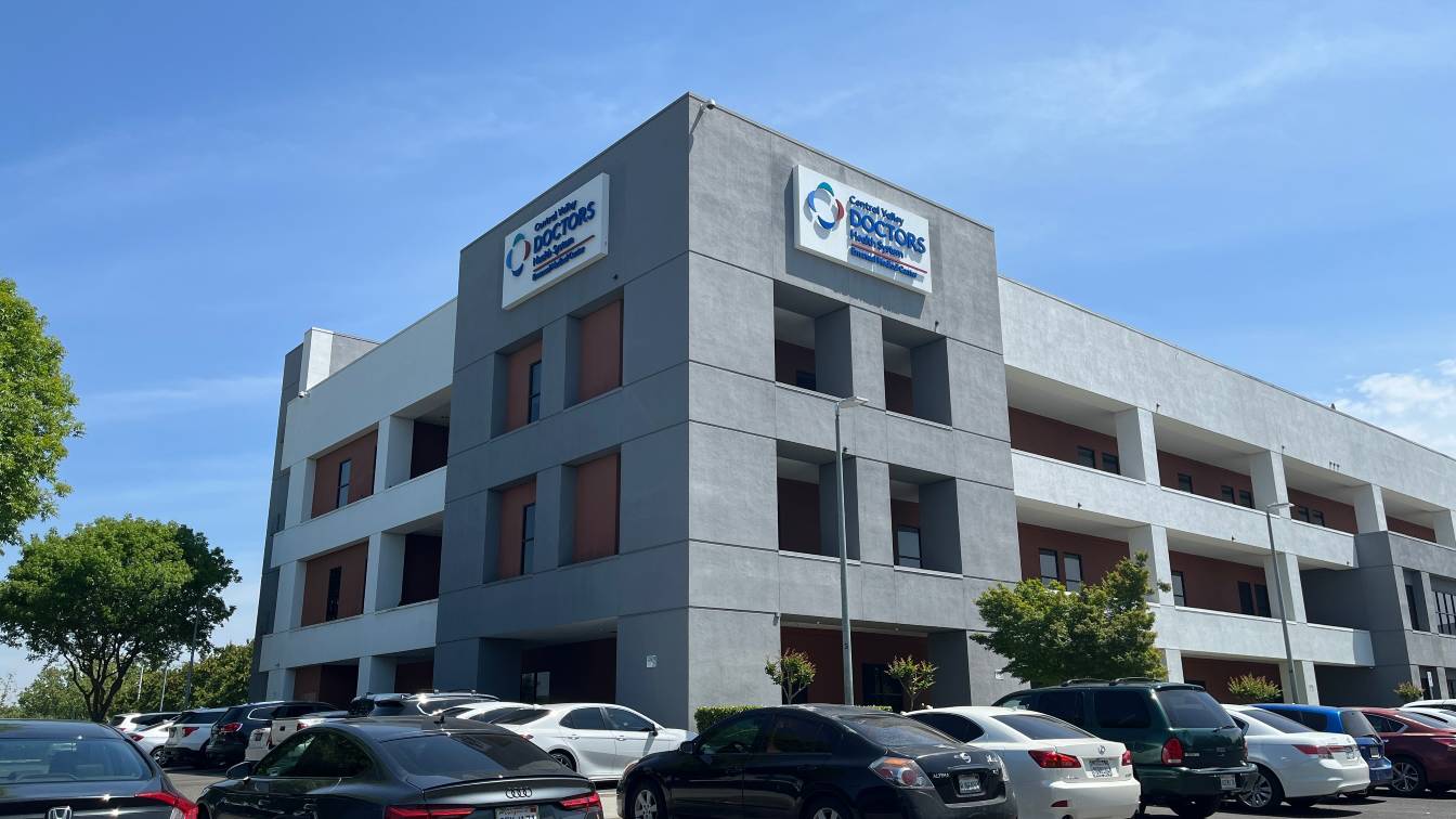

Emanuel Medical Center - Outpatient Imaging

Modesto Radiology Imaging

Ruby E. Bergman Women's Diagnostic Center
Diagnostic Imaging Services in Central Valley (may vary by location)
- Biopsies – involve taking a sample of body tissue for an examination under a microscope.
- Bone densitometry (DEXA) – uses low-energy X-rays to help doctors scan a patient’s forearm, hip and lower back.
- Breast Imaging - ACR Breast Centers of Excellence, a trusted facility that has proven its commitment to delivering the highest quality, most coordinated, and patient-focused breast cancer care available, backed by rigorous standards and expert teams.
- 3D Tomosynthesis– uses special types of X-ray to create a detailed view of the breast and help doctors detect breast cancer.
- Stereotactic Breast Biopsy
- Breast Ultrasound
- Biopsy and Breast Cyst Aspiration
- CT/CTA scan – uses computers and rotating X-ray machines to capture cross-sectional images of various body parts and internal organs such as the brain, abdomen, chest, head, heart knee, shoulders and spine. A CT scan can also create images of the bones, blood vessels and soft tissues. A CTA uses CT scan and the injection of a dye to create images of blood vessels in the neck and head.
- CT Perfusion - uses CT scan and the injection of dye to create images of the brain tissue.
- Digital X-ray – a modern X-ray variant that uses 80% less radiation than the traditional X-ray in creating images of the body’s internal organs and bones.
- Fluoroscopy – like an X-ray movie or an X-ray in motion that captures moving body structures in detail to help diagnose various types of illnesses.
- Magnetic resonance imaging (MRI) – uses an electromagnet, radio waves and a computer to produce detailed images of internal organs and body parts. Doctors commonly request an MRI to get a detailed view of the brain, abdomen, bones, spine, pelvis and joints.
- MR angiography – a detailed MRI of the blood vessels that helps diagnose diseases in the blood vessels supplying the brain, kidneys, legs and lungs.
- MR Perfusion - a detailed MRI of the brain tissue that helps diagnose diseases in the brain.
- Nuclear Medicine - shows how organs function using injected radioactive tracers, revealing biological processes (like blood flow or metabolism) with high sensitivity, while a CT scan uses external X-rays to show detailed anatomy (structures and shapes
- Positron emission tomography (PET)/CT scan – uses both PET and CT in one machine. This type of exam gives doctors more information about the structure and function of cells and tissues in the body.
- Ultrasound – creates images of internal organs using high frequency sound waves. e offer various types of ultrasound such as abdominal, pelvic, obstetrical and vascular ultrasound.
| Services | DHM | MIC | DMC | MRI | EMC | RB |
| Bone density/DEXA | X | X | X | |||
| CT Scan / CTA | X | X | X | X | X | |
| Fluoroscopy | X | X | X | X | ||
| Interventional radiology treatment | X | X | X | |||
| Mammography | X | X | X | |||
| 2D digital mammograph | X | X | X | |||
| 3D tomosynthesis | X | X | X | |||
| Stereotactic breast biopsy | X | X | X | |||
| Wire localization procedure | X | X | X | |||
| MRI | X | X | X | X | ||
| Breast MRI | X | X | ||||
| Neuro interventional radiology | X | |||||
| Open MRI | X | X | ||||
| Nuclear medicine | X | X | X | |||
| PET/CT | X | X | X | |||
| Ultrasound | X | X | X | X | X | |
| Breast ultrasound | X | X | X | |||
| Biopsy & breast cyst aspiration | X | X | X | |||
| X-ray (digital) | X | X | X | X | X | |
| Manteca | ||||||
| DHM = Doctors Hospital of Manteca | ||||||
| MIC = Manteca Imaging Center | ||||||
| Modesto | ||||||
| DMC = Doctors Medical Center | ||||||
| MRI = Modesto Radiology Imaging | ||||||
| Turlock | ||||||
| EMC = Emanuel Medical Center | ||||||
| RB = Ruby E. Bergman |
Advanced Imaging Treatment Solutions
- Interventional Radiology and Neuro Interventional Radiology
- Chemoembolization – delivers cancer treatment directly to the tumor through the blood supply.
- Embolectomy - removal of blood clots, noninvasive treatment for acute ischemic stroke.
- Embolization – blocks abnormal blood flow or abnormal organ function, such as:
- Uterine artery embolization to treat fibroid tumors in the uterus.
- Arterial embolization of ruptured and unruptured aneurysms and brain bleeds.
- Radiofrequency ablation – enables more precise cancer treatment.
- Thrombolysis – helps dissolve blood clots.